Visual cortex
| Brain: Visual cortex | |
|---|---|
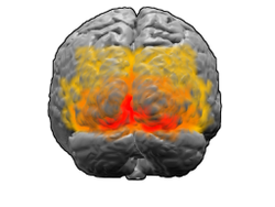 An orange, 19 fields are shown with yellow red, 18 fields 17 fields (primary visual cortex) in the brain map of Brodmann. | |
| Name | |
| Japanese | Visual cortex |
| English | Visual area, Visual cortex |
| Structure concerned | |
| Higher structure | Cerebral cortex, occipital lobe |
| Related information | |
| Brede Database | Hierarchy relations, coordinate information |
| MeSH | Visual+Cortex |
Primary visual cortex (or with line cortex (striate cortex, the cable broadcasting cortex)) that the term visual cortex (as soon as I do it and write it British: visual cortex) is abbreviated to V1 And I show V2, V3, V4, outer stripe cortex (with extrastriate cortex, cortex out of the cable broadcasting, the cable broadcasting outside of the territory cortex) abbreviated to V5. The primary visual cortex is equal to 17 fields in the brain map of Brodmann anatomically.
Table of contents
Introduction
Primary visual cortex (V1) is granule cortex (Koniocortex) located around calcarine sulcus in the occipital lobe and receives direct information from outside knee-formed nucleus.

V1 conveys information on two kinds of main sight courses called a back side cortex visual pathway and the ventral cortex visual pathway.
- The back side cortex visual pathway begins in V1 and, via V2, back inside field, the MT field (with V5), goes to the occiput top cortex. The back side cortex visual pathway is sometimes called the where course and is connected with saccade and reaching movement using exercise, the control of a position and eyes and the arm of the object particularly the sight information [1].
- The ventral cortex visual pathway begins in V1 and sits at the lower both sides of the head cortex via V2, V4. I am sometimes called the what course, and the ventral cortex sight course is related to the representation (picture amounting to consciousness) of recognition and the shape for the sight. In addition, I am related to the storage of the long-term memory.
Dichotomy (or dichotomy of where/what, dichotomy of action (action)/ perception (perception)) of a back side cortex visual pathway and the ventral cortex visual pathway [1] is defined as Ann garfish rider (Ungerleider) first by ミシュキン (Mishkin) [2] and sparks a debate among an optic nerve scientist and psychologists now. Probably this dichotomy simplifies the real event with the visual cortex too much. This study is based on discovery that a judgment of the perception is twisted by Ebbinghaus optical illusion, but being warped of perceiving it does not usually occur when a person reacts by the actions such as grasp campaigns. However, it is said that an illusion is taking place by these optical illusion as for the action and the perception in the recent study [3] equally.
When sight stimulation was exhibited by a receptive field, the neuron of the visual cortex produces action potential. From a definition, the receptive field points at a thing causing action potential in all fields of vision. However, about a certain neuron, I reply to the subset of specific stimulation of the receptive field of the neuron. This characteristic is called tuning. Simpler tuning is carried out with the earlier visual cortex. For example, the neuron with V1 catches fire for the perpendicular flight that the receptive field shows. More complicated tuning is carried out with a more highly advanced visual cortex. For example, the neuron with the lower both sides of the head cortex (IT) catches fire for a specific face emerging to the receptive field.
The visual cortex catches the supply of the blood from the calcarine branch of the posterior cerebral artery most.
Recent study
I measure action potential by the electrode which I stabbed a cat, a ferret, a rat, a mouse, the brain of the monkey with in the study of the primary visual cortex, and there is a thing measuring fMRI signal of V1 of a thing and Homo sapiens and the monkey which measured the endogenous optics signal in the animal.
By the discovery with these days, as for the signal measured by fMRI of V1 of the Homo sapiens, there is a thing to strongly receive adjustment (attentional modulation) by the attention. This result is in contrast to the result that a change was hardly seen in for the ignition of the neuron by adjustment by the attention in the physiologic study of the macaque monkey. However, the study of the macaque monkey checked the spikes activity of 1 neuron, but the nerve base of the fMRI signal comes close to postsynaptic reinforcement (PSP) mainly. Therefore, the difference in this result is not the thing which just means physiologic difference between Homo sapiens and マカク.
In the study of other recent V1, I completely explain a characteristic of the tuning, and there is the thing which tried it to use it as a model of the cortical カノニカル circuit.
The damage of the primary visual cortex produces blind spots ("a hole" made in the field of vision). Interestingly, I can use the sight information in the blind spot though the patient having a blind spot cannot perceive it consciously. This phenomenon is called blindsight, and it is studied widely by the researcher who is interested in brain activity (neural correlate of consciousness) that related to consciousness.
Primary visual cortex (V1)
It is a domain checked with the visual cortex of the primary visual cortex brain best. In all studied mammals, the primary visual cortex is located in the occipital pole of the occipital lobe (the occipital lobe takes handling of sight stimulation). It is the simplest, and the primary visual cortex specializes it in processing of the information about standstill or the exercising object with a visual cortex active earliest and shows power for pattern recognition again.
The domain called the primary visual cortex defined functionally is quite equal to defined line cortex anatomically. "Line cortex (striate cortex) As for the name called ", even the axonal naked eye made a lengthening myelin sheath from the lateral geniculate body called the Gen Nari line (stria of Gennari) to the IV layer of the gray matter comes from an identifiable big striped pattern.
The primary visual cortex is divided into six levels of layers different functionally labelled from the I layer to the VI layer. The IV layer catching the input of most sight information from inner lateral geniculate body (LGN) of 4A, 4B, 4Cα, 4Cβ is divided into four levels more. The 4Cα sub layer receives most giant cell-related input from a lateral geniculate body, and the 4Cβ layer catches the input from the parvocellular course.
The mean neuronal number of primary visual cortexes of the human adult is estimated as approximately 140 million because of each cerebral hemisphere. (Leuba & Kraftsik, Anatomy and Embryology, 1994)
Function


V1 has a clear map of the space information in the sight. For example, the calcarine sulcus superior wall of the Homo sapiens strongly replies to the sight information for the lower half of the field of vision, and the lower wall replies to half on a field of vision. V1 from retina can consider the mapping of this レチノトピー to be the conversion of the sight image conceptually. A field of vision and the correspondence of the position in V1 are very accurate, and even the blind spot is mapped with respect in V1. This correspondency is basic evolutionarily and is seen in most animals having V1. In an animal and the Homo sapiens having central fovea, a domain accounting for a big ratio of V1 maps a small domain in the center of the field of vision with respect. This phenomenon is known as cortex expansion (cortical magnification). Probably, for accurate spatial coding, the size of the receptive field of the neuron of V1 will be smallest in the microdomain of all visual cortexes.
The tuning properties of the V1 neuron (reply selectivity of the neuron) greatly vary according to time. In (before and after 40ms), each V1 neuron is strongly tuned up for a short time for a small group of the stimulation. In other words, I can divide the responsiveness of each neuron into slight difference of a direction of the sight stimulation, space frequency and the color. Furthermore, with Homo sapiens, I look at the both eyes, and each V1 neuron of the をする animal tunes it up for one of both eyes with eyes superiority literally. I gather, and the neuron of V1 and the general primary sensory area makes the structure called the cortex column with a thing with similar tuning properties. David Hugh bell and torr Sten ウィーセル proposed a classic model (the classic ice-cube organization model) about the cortex column of eyes superiority and two tuning properties of the direction. However, this model did not fit about many other characteristics that neurons such as color or the space frequency were tuned up. The accurate constitution of the cortex column of all these V1 is a still hot topic in a recent study.
It is thought that the early reply of the V1 neuron is comprised of a filter (spatiotemporal filters) between the selective space-time now. In the space dimension, as for the function of V1, much; it is thought that is local, and resembled complex Fourier transform spatially. Such a filter can take a neurologic step about space frequency and a direction, exercise, a moving direction, speed (as for therefore time frequency) and the characteristic of many other space-time intervals theoretically. The experiment in the V1 neuron demonstrates this theory, but produces the newer question.
The neuron of V1 replies to wider structure of scene (scene) late in terms of time in (after 100 ms) (Lamme & Roelfsema, 2000). It is thought that such the reply properties occur because of recurrent processing (influence from the highly advanced domain to the domain of the low level) and the horizontal combination (Hupe et al 1998) of the cone cell.
The sight information relayed to V1 is encoded as local contrast as the image which is spatial (or optical). For example, the line where black, the other side divide the black and white that the contrast that the image consisting of the white is local is biggest into is encoded, and one side encodes only the neuron that there is a little information (blackness and innocence in itself) of the brightness. As information is relayed to the highly advanced sight domain, nonlocal-like frequency / phase signal is encoded more. In the early days of the sight processing with such cortex, the spatial position of the sight information is strong in coding of the local contrast, and an important thing is a saved point.
V2
V2 is called the previous cable broadcasting cortex (prestriate cortex) [4] and, in the second largest domain of the visual cortex, is a domain at the first beginning of the sight association area. This domain receives strong feedforward connection from V1 and sends connection to V3, V4, V5. In addition, I have strong feedback connection to V1.
V2 is divided into the back side of the right and left hemisphere and four quadrants of the ventral anatomically. These 4 domains form a complete map of the sight world together. V2 has V1 and many common characteristics functionally. The cell is tuned up by the simple characteristics such as a direction or space frequency, the color. As for many replies of the V2 neuron, the direction of the subjective outline and the sight stimulation receive adjustment by a part of the figure or a part of the ground or a more complicated characteristic. (Qiu and von der Heydt, 2005).
The cell of V2 has a small adjustment (attentional modulation) by the attention (it is bigger than V1 and is smaller than V4), and, by a recent study, what is tuned up by a pattern complicated moderately by reacting to the plural directions of the different low rank domain in the single receptive field again becomes clear.
The third visual cortex complex including V3
The term third visual cortex complex shows a cortex domain contacting with the front part of V2 and includes V3 domain. In Homo sapiens, as for the name called "complex", in the domain in front of V2, a debate depends on a fact to be up by a researcher to include two or three functional low-ranking domains about the exact range of the domain called V3. For example, there is a domain called "back side V3"(dorsal V3) in the upper part of the cerebral hemisphere and proposes デイヴィット station wagon Essen (David Van Essen) and others (1986) if different from" ventral V3" (posterior ventral V3, ventral domain, ventral posterior area, VP) located in the retainer of the brain. Ventral V3 receives different connection from the different domain of the brain, and is different from back side V3 by many stainings; is dyed, and do, and the sight stimulation is different; put it together, and reply to (e.g., there are many color selective neurons in ventral V3). As other low rank domains, V3A and V3B are reported in Homo sapiens. These low rank domains exist near back side V3, but are distinguished from V2.
Back side V3 usually of the back side cortex visual pathway it is thought partly, and receive input from V2 and a primary visual cortex, and project it on posterior parietal cortex. It is located in 19 fields in the brain map of Brodmann anatomically. That the V3/V3A domain is concerned with handling of broad-based movement by a recent study by fMRI [5]. Other studies to think about partly of the big domain called back inside field (DM) expressing all fields of vision in back side V3 exist. The neuron of the back inside field replies to the movement that synchronized of the big pattern to occupy most of the fields of vision. (Lui and collaborators, 2006)
Ventral V3 (VP) has the very weak combination from a primary visual cortex and strong combination with the inferior temporal gyrus. In the early study, ventral V3 set out only for the domain that expressed (the upper part of the fixation point) for the first half of the field of vision. しかし、より最近の研究では、腹側 V3 は以前考えられていたよりもより広く、他の視覚野と同様に視野全体を表現する領域を考えられている。 この改訂されたより広い腹側 V3 はローサ (Rosa) とツィーデル (Tweedale) によって腹外側後部領域 (VLP) と呼ばれた[6]。
V4
V4はマカクザルでは、有線外視覚皮質に存在する視覚野の一部である。V4 は V2 の前部に位置し、後下側頭野 (PIT) の後部に位置する。V4 は左右のV4d、V4vの4領域から成るとされ、さらに尾側と吻側の下位領域を加える者もいる。ヒトにおける V4 ホモログはまだ知られておらず、精査されている事柄である。
V4 は腹側皮質視覚路の3番目の領域で、V2 からの強いフィードフォワードな入力を受け, 後下側頭野 (PIT) へと強い結合を持つ。また V1 からの直接の入力をその中心部へ受けている。加えて、 V5 と背側前月状回 (DP) へと弱い結合が存在する。
V4 は腹側皮質視覚路において強い注意による調節 (attentional modulation) を受ける最初の領域である。多くの研究では、選択的注意が V4 の発火頻度を20%変化させるとしている。モラン (Moran) とデシモーネ (Desimone) による、影響力の大きい論文は、このような効果を特徴付け、注意による効果は視覚野のどこにおいても見られることを明らかにした最初の論文である[1][7]。
V1 のように V4 は方向、空間周波数、色にチューンしているが、V1 とは異なり、V4 は対象の特徴の中間的な複合性、例えば、単純な幾何学的形状などに、チューンしている。しかし、V4 のチューニング空間の完全なパラメーターの記述を行った者はいない。また、V4 は下側頭回のように顔などの複雑な対象にはチューニングされていない。
V4 の発火特性は1970年代後半にこの領域の名付け親でもあるセミール・ゼキ (Semir Zeki) によって初めて描写された。それまでは、 V4 はその解剖学的な名である前月状回として知られていた。元々、ゼキは V4 の目的は色情報を処理するためであると考えていたが、1980年代前半に V4 は他のより初期の視覚野と同様に形の認知に直接関わっていると示された。この研究はアンガーライダー (Ungerleider) とミシュキン (Mishkin) により1982年に初めて提唱された2経路仮説 (上述) によっても支持されている。
最近の研究によって、V4 は刺激の顕著性(salience)をコードする長期可塑性を示し、その可塑性は前頭眼野からの信号によってコントロールされることが分かった。これにより、注意によって受容野の空間的な特性が変化する。
V5/MT野
V5はMT野 (middle temporal) としても知られ、外線条野の一部であり、運動の知覚、局所運動信号のグローバルな知覚への統合、及びある種の眼球運動における主要な役割を担うと考えられている[8]。
神経結合
MT 野は様々な皮質及び皮質下の脳領域に結合している。その入力は V1、V2、背側 V3、背内側野[9] [10]、 外側膝状体の顆粒細胞(koniocellular)領域[11]、及び、下視床枕から受けている。MT 野への投射のパターンは視野の中心と周辺で幾分異なっている。後者では、皮質正中部と脳梁膨大後部からの入力を受ける[12]。
一般的には、MT 野への"最も重要な"入力はV1に由来すると考えられている[8]。しかし、いくつかの研究は MT 野のニューロンは V1 ニューロンが破壊または不活性化された後でも、視覚情報に対して、多くの場合は方向選択的に応答できることを示した[13]。加えて、セキール・ゼキ (Semir Zeki) らによる研究ではある種の視覚情報は V1 に到着する前に MT 野に到着することが示唆されている。
MT 野はその主な出力を、 FST や MST、V4t (中側頭半月部) を含むその領域を直接取り囲む領域へと送っている。他の MT 野の出力先は眼球運動に関係する前頭葉と頭頂葉の領域 (前頭眼野と外側頭頂間野(LIP))である。
Function
MT 野のニューロンの電気生理学的な特性を調べた最初の研究により、大部分の細胞が運動する視覚刺激の速度と方向にチューニングされていることが分かった[14] [15]。このような結果は MT 野が視覚運動の処理に重要な役割を担っていることを示唆している。
MT 野の損傷による研究によっても、その運動知覚と眼球運動における役割は示されている。また、運動を知覚することが出来ず、世界が静的な"フレーム"の連続に感じられる患者の神経心理学的研究により、霊長類における MT 野のヒトにおけるホモログは V5 であると分かった[16][17]。
しかし、V1 のニューロンも運動の方向や速度にチューニングされていることから、これらの初期の結果は、V1 では処理不可能な何を MT 野では処理可能なのかという問題をまだ解けてはいない。多くの研究によって、この領域が局所的な視覚運動信号を複雑な対象の大局的運動(global motion)へと統合していることが分かった[18]。 例えば V5 の損傷により、運動の知覚と複雑な刺激の処理に障害が起きる。V5 には複雑な視覚的特徴 (線の端やコーナーなど) の運動に選択的に応答する多くのニューロンがある。 V5 にあるニューロンの微小刺激は動きの知覚に影響を及ぼす。例えば、上向きの運動に選択的に応答するニューロンを見つけて、電極により刺激したとき、ニューロンを刺激されたサルは上向きの運動をより多く報告しやすくなる[19]。
MT 野で行われている計算の正確な形に関してはいまだに論争が存在する[20]。そして、ある研究では、特徴の運動は V1 のような視覚野における低次の領域でも、実はすでに利用可能であるとしている[21] [22]。
機能的な構成
MT 野は方向コラムにより構成されていることが示されている[23]。デアンジェリス (DeAngelis) は MT 野のニューロンは両眼視差へのチューニングに基づいて構成されていると主張している[24]。
参考文献
- ^ a b Goodale & Milner (1992). "Separate pathways for perception and action.". Trends in Neuroscience 15: 20-25. doi:10.1016/0166-2236(92)90344-8.
- ^ Ungerleider and Mishkin (1982). Ingle DJ, Goodale MA and Mansfield RJW. ed. Analysis of Visual Behavior. MIT Press.
- ^ Franz VH, Scharnowski F, Gegenfurtner (2005). "Illusion effects on grasping are temporally constant not dynamic.". J Exp Psychol Hum Percept Perform. 31(6): 1359-78.
- ^ Gazzaniga, Ivry & Mangun: Cognitive neuroscience, 2002
- ^ Braddick, OJ, O'Brian, JMD, et al (2001). "Brain areas sensitive to visual motion.". Perception 30: 61-72. doi:10.1068/p3048.
- ^ Rosa MG, Tweedale R (2000) Visual areas in lateral and ventral extrastriate cortices of the marmoset monkey. J Comp Neurol 422:621-51.
- ^ Moran & Desimone. Selective Attention Gates Visual Processing in the Extrastriate Cortex. Science 229(4715), 1985.
- ^ a b Born R, Bradley D. "Structure and function of visual area MT.". Annu Rev Neurosci 28: 157-189. PMID 16022593.
- ^ Felleman D, Van Essen D. "Distributed hierarchical processing in the primate cerebral cortex.". Cereb Cortex 1 (1): 1-47. PMID 1822724.
- ^ Ungerleider L, Desimone R (1986). "Cortical connections of visual area MT in the macaque.". J Comp Neurol 248 (2): 190-222. doi:10.1002/cne.902480204. PMID 3722458.
- ^ Sincich L, Park K, Wohlgemuth M, Horton J (2004). "Bypassing V1: a direct geniculate input to area MT.". Nat Neurosci 7 (10): 1123-8. doi:10.1038/nn1318. PMID 15378066.
- ^ Palmer SM, Rosa MG (2006). "A distinct anatomical network of cortical areas for analysis of motion in far peripheral vision.". Eur J Neurosci 24(8): 2389-405.
- ^ Rodman HR, Gross CG, Albright TD (1989) Afferent basis of visual response properties in area MT of the macaque. I. Effects of striate cortex removal. J Neurosci 9(6):2033-50.
- ^ Dubner R, Zeki S (1971). "Response properties and receptive fields of cells in an anatomically defined region of the superior temporal sulcus in the monkey.". Brain Res 35 (2): 528-32. doi:10.1016/0006-8993(71)90494-X. PMID 5002708..
- ^ Maunsell J, Van Essen D (1983). "Functional properties of neurons in middle temporal visual area of the macaque monkey. I. Selectivity for stimulus direction, speed, and orientation.". J Neurophysiol 49 (5): 1127-47. PMID 6864242.
- ^ Hess, Baker, Zihl (1989). "The" motion-blind" patient: low-level spatial and temporal filters". Journal of Neuroscience 9 (5): 1628-1640.
- ^ Baker, Hess, Zihl (1991). "Residual motion perception in a" motion-blind" patient, assessed with limited-lifetime random dot stimuli". Journal of Neuroscience 11 (2): 454-461.
- ^ Movshon, J.A., Adelson, E.H., Gizzi, M.S., & Newsome, W.T. (1985). The analysis of moving visual patterns. In: C. Chagas, R. Gattass, & C. Gross (Eds.), Pattern recognition mechanisms (pp. 117-151), Rome: Vatican Press.
- ^ Britten & Van Wezel 1998
- ^ Wilson, H.R., Ferrera, V.P., & Yo, C. (1992). A psychophysically motivated model for two-dimensional motion perception. Vis Neurosci, 9 (1), 79-97.
- ^ Tinsley, C.J., Webb, B.S., Barraclough, N.E., Vincent, C.J., Parker, A., & Derrington, A.M. (2003). The nature of V1 neural responses to 2D moving patterns depends on receptive-field structure in the marmoset monkey. J Neurophysiol, 90 (2), 930-937.
- ^ Pack & Born, 2003
- ^ Albright T (1984). "Direction and orientation selectivity of neurons in visual area MT of the macaque.". J Neurophysiol 52 (6): 1106-30. PMID 6520628.
- ^ DeAngelis G, Newsome W (1999). "Organization of disparity-selective neurons in macaque area MT.". J Neurosci 19 (4): 1398-415. PMID 9952417.
関連文献
日本語のオープンアクセス文献
- 佐藤宏道, 勝山成美 「一次視覚野の機能構築とその役割」 日本視能訓練士協会誌 Vol.21 (1993) pp.154-160
- 乾 敏郎 「視覚の計算理論とモジュールの統合」 計測と制御 Vol.33, No.3 (1994) pp.222-226
関連項目
外部リンク
- 第一次視覚野が壊れると見えなくなる - 中部学院大学 三上章允
- サルやヒトの脳には沢山の視覚野がある - 中部学院大学 三上章允
- ユタ大学の(Matthew Schmolesky) による一次視覚野の解説
- ハーバード大学のデービット・ヒューベルによる視覚野の構造の解説
- 視覚の計算モデリングのシミュレーター
- (百科事典)「Area V1」 - スカラーペディアにある「一次視覚野」についての項目(英語)
- 眼優位性 - 脳科学辞典
This article is taken from the Japanese Wikipedia Visual cortex
This article is distributed by cc-by-sa or GFDL license in accordance with the provisions of Wikipedia.
In addition, Tranpedia is simply not responsible for any show is only by translating the writings of foreign licenses that are compatible with CC-BY-SA license information.
0 개의 댓글:
댓글 쓰기