White blood cell

I point to the immune charge cell about living body defense in a leukocyte (はっけっきゅう British: White blood cell or British: Leukocyte) wide sense. However, besides, an immune charge cell (include polymorph, monocyte, the dendritic cell which did including neutrophils, and do not include a lymphocyte) differentiating from a pro-cell ingredient and marrow included in the blood outrider cell may express neutrophils in a narrow sense alone [1]; collect it, and it is necessary to point to what by context, or to note the thing that may be different at all. In general, this clause is included in blood mainly to be considered to be a generic name of a lymphocyte, polymorph, the monocyte and is based on a definition called one of the cell ingredients detected by general inspection. This cell ingredient is a cell derived from the hematopoietic stem cells which assume the exclusion of the alien substance including bacteria, the virus which invaded the body from the outside and the exclusion of the cell which finished a neoplastic cell, a duty a role.
I am often referred by the blood tests a WBC.
The size from 6 30µm (as for the macrophage more than it). There is not the gender gap, and the number is around 9,500 from 3500 per normal blood 1µL (I refer to the standard value melody of the white blood-cell count).
Table of contents
Kind of the white blood cell
There is polymorph, a lymphocyte, monocyte in the peripheral blood; the polymorph of the dyeing by the Giemsa's stain is done, and is classified in neutrophils, acidophile, three of the basophil by a difference of the [2].
Therefore, white blood cells in the peripheral blood are usually assumed five kinds of neutrophils, acidophile, basophil, a lymphocyte, the monocyte.
The polymorph is produced with marrow and occupies 3/4 degree from half of the white blood cell in the peripheral blood [3]. There is a granule having disinfection to cytoplasm. The lymphocyte occupies 40% from 20 in the peripheral blood, and the monocyte occupies approximately 6% from 3 [3].
Differentiation of the monocyte fits every progress organization in the organization, and various macrophages playing a role such as the somatic processing that it was for bookmaking food of the alien substance, an unnecessary, release of Ag presenting cytokine to a humoral immunity cell exist.
Standard value of the white blood-cell count
The standard value of the white blood cell is considered to be 3500 - 9500/µL with the representative textbook of the internal medicine [2]. However, the white blood-cell count in the peripheral blood is highly individual and fluctuates among one person in a short time again. Therefore there is a difference for the setting of the standard value every every inspection organization or 成書, and the standard lower limit level varies in 3300 - 4000/µL, the standard upper limit level in the adults with 8000 - 11000/µL [3]. A newborn baby and infants have more numbers than an adult, and the thing more than 20000/µL is not rare in newborn babies, too. In addition, the ratio of lymphocyte is high in swaddling bands and becomes around 70% from a newborn baby [3].
When there are diseases, I increase than the standard value and decrease [4].
Than the standard value in the case of increase white polycythemia (Leukocytosis)
- More than 50,000/µL
- Leukemia, myeloproliferative disease, chestnut grain tuberculosis, sepsis, malignant tumor
- 10,000 - 50,000/µL
- Infectious disease, autoimmune disease, metabolism disorder, drug poisoning, leukemia, myeloproliferative disease, influence, stress, smoking, the pregnancy of the steroid
Than the standard value in the case of decrease leukopenia (Leukopenia)
- 1,000 - 3,000/µL
- Aplastic anemia, side effect, drug allergy of an anticancer agent, the radiation therapy, metastasis of cancer to marrow, marrow dysplasia syndrome, pernicious anemia, 脾機能亢進, typhoid fever, virus infectious disease, osteomyelofibrosis, myxedema, AIDS, agranulocytosis
- Less than 1,000/µL
- It is the same as 1,000 - 3,000/µL
The increase and decrease of the white blood-cell count includes the diseases mentioned above, but cannot make a definite diagnosis only with a white blood-cell count [5].
Neutrophilic

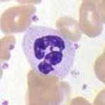
Neutrophilic (neutrophile white blood cell) (Neutrophil) occupies 70% from 50 of the leukocyte whole in the peripheral blood [3] and occupies 95% from approximately 90 in the polymorph. Neutrophils deal mainly, but neutrophils gather for the infection such as bacteria or the fungus first and the neutrophils do not perform the antigen presentation to a humoral immunity cell. Macrophages englobe the alien substances such as the bacteria which neutrophils were not able to process and perform the antigen presentation and get humoral immunity. The pus which occurs from a wound after having done injuries with bacteria rustle, and is mainly with the body of dead neutrophils.
Shape
Of the colorlessness semitransparency is almost spherical, but the form is not decided because take out a pseudopodium, and carry out an ameboid activity flourishingly. Having a germicidal special granule dyed a neutral pigment by the Giemsa's stain that is standard hemocyte dyeing, I may be said to be a many nuclei white blood cell in one (leaf) of which a nucleus is divided into when it matures. The neutrophils of the last completion form are called share leaf karyosphere, and the nucleus is divided, but is connected by the linin between the nucleus. With the stage when a nucleus is big before becoming the share leaf karyosphere, and bent jelly beans is bacillary (bacillary karyosphere). It is a lysosomal kind, and the germicidal granule is made with a Goldi body (net device). The diameter is 12 - 15µm [6] and is bigger than a lymphocyte in the white blood cell and is smaller than monocyte, a macrophage.
Amount, life
The neutrophils from 2000 to around 7,500 per 1 microliter are included in the peripheral blood [3], and neutrophils of the order (figure) of 10 10 乗個 almost exist in the peripheral blood of the adult. In the case of 50 kg in weight, it is the amount from about 8 billion to around 30 billion.
However, as for the neutrophils, neutrophils of the quantity to be equal to a vascular wall and an organization, spleen, liver in peripheral blood exist as a border pool. Furthermore, there is a retention pool of the quantity of 30 times to marrow from 10 in the peripheral blood, and order of 10 11 乗, the neutrophils of hundreds of billions of figures exist in all in the living body.
Because there is a big retention pool, the neutrophils in the retention pool are mobilized for at the time of bacterial infection, and the neutrophilic numbers in the peripheral blood increase immediately. In addition, it is the change of a few bodies such as a meal and exercise, the stress, but the neutrophilic number is easy to change into a vascular wall by a change of the bloodstream because the neutrophils which existed move to a border pool in peripheral blood by stay. At the time of the bacterial infection, the production in the marrow is aggravated by a function of inflammatory cytokine, too.
When there is not infection, some neutrophils move from blood vessel in an organization, and they exist.
The life of neutrophils in the blood is almost considered to be approximately ten hours for less than one day. It is a few days in the organization.
The neutrophils are produced in marrow, but are made 10 11 乗個 (100 billion) degree per day [3].
Structure of the living body defense of neutrophils
When bacteria infect the living body, the neutrophils migrate it in the infected inflammatory part and englobe a meeting, bacteria and sterilize it.
I migrate it and it

By the tissue which bacteria and a fungal kind invaded, an interstitial macrophage and obese cell react promptly and release cytokine such as interleukin-1 (IL-1), and, by those cytokine, the interstitial cell wakes up an inflammatory change. In addition, including other process, the organization which caused an inflammatory change releases the various kinds of neutrophils migration stimulating factors of chemokines (cytokine) and others doing interleukin-8 (IL-8) with a representative various kinds of. The neutrophils which took in the material which those stimulating factors and bacteria oneself produce, an activated complement with a surface receptor let you activate migration exercise. The speed becomes up to 40μm/min. The neutrophils migrate it with the receptor which the surface has a lot in the dark thin いを feeling collecting, dark direction of the density of the factor of the density of the stimulating factor and gather it to an infection nest. In many cases, the infection nest is out of blood vessel, and the neutrophils must pass a vascular wall. The neutrophils adhere to a blood vessel epithelium in a capillary wall near an irritation, and a blood vessel epithelium cell and a lot of each neutrophilic wake up a change by a factor, and the neutrophils pass through a blood vessel epithelium cell. The neutrophils which appeared outside blood vessel migrate the organization and arrive at the infection den.
The neutrophils which there is in retention pools in the marrow are stimulated and start migration exercise, and the production of neutrophils is aggravated with the marrow again by the migration stimulating factor from the inflammatory organization. A large quantity of neutrophils will be mobilized for the infection of the bacteria by them.
A property to move by a leukocyte migration stimulating factor (ロイコエグレシン, Roy cocaine, lymphokine, bacterial toxin, resolution product of the complement) to occur because of inflammation in this way is called chemotactic (chemotaxis).
I englobe it and sterilize it and it
The neutrophils which arrived at the infection den englobe it from the contact to a bacteria and sterilize the bacteria which I swallowed.
I recognize that the neutrophils contact a bacteria with an alien substance through a surface receptor and adhere and bond it. I take in a connected alien substance in neutrophils as if a neutrophilic form and essence film giving this.
The bacteria taken in in neutrophils is sterilized by three means. One produces active oxygen and hydrogen peroxide, hypochlorous acid by oxygen-based work and sterilizes it. The other sterilizes it with hydrolases released by a granule. In late years form a net of the chromatin called NETs(neutrophil extracellular traps), and is known to catch a microbe [7]; [8].
It is processed whether the neutrophils which swallowed bacteria die before long and the body becomes the pus and is released outside a body by interstitial macrophages.
Refer to a neutrophilic item for a process of the living body defense in detail.

I it in a differentiation process
Including neutrophils, all blood corpuscles come from existing hematopoietic stem cells in marrow. The hematopoietic stem cells differentiate to red blood cell various white blood cells, platelets in marrow, but differentiation matures in hematopoietic stem cells, marrow system stem cell (marrow system outrider cell), polymorph, monocyte system outrider cell, polymorph outrider cell, myeloblast, the former myelocyte, myelocyte, back myelocytic order when I finally differentiate to neutrophils. Furthermore, I differentiate to share leaf karyosphere through bacillary karyosphere, but call it neutrophils with two of this last.

I push forward a direction of the differentiation little by little while the cell which is divided from hematopoietic stem cells, and has begun to differentiate being divided flourishingly, and increasing numbers. It is the morphologic observation with the microscope and is finally the cell which pro-the polymorph such as neutrophils, differentiates, or, until an outrider cell, a myeloblastic stage, the identification is difficult, but a granule begins to produce it from a myeloblastic stage and comes to be able to judge it with a pro-polymorph cell from a stem cell morphologically. When it is a promyelocytic stage, a tendency to differentiation to neutrophils becomes clear.
A primary granule (アズール granule) begins to arise from a myeloblastic stage and comes to have an abundant primary granule (アズール granule) with the previous myelocyte. I lose my eyesight of the primary granule at the myelocytic stage, and the second granule (special granule) emerges as a substitute for (I do not see it, but exist). Furthermore, there come to be various germicidal granules to the neutrophils including the third granule.
I wake up approximately two times of cell division with myelocyte and, polymorph system and the stage when I became able to judge it later, add to a number with previous myelocyte with a myeloblast twice once. When it is back myelocytic stage, the ability to divide the cells is lost. Sometimes write the time of approximately 11 days at the stage after the myeloblast and usually mature.
At the immature stage including a myeloblast and the previous myelocyte, the nucleus of the cell is big, and the structure (chromatin structure) in the nucleus is delicate in a circle, but the nucleus becomes distorted small so that differentiation, maturity advances, and the structure becomes coarse. When it is a stage called "the bacillary karyosphere" which is the jelly beans form that a nucleus was warped, I am recognized with finished neutrophils, but maturity advances more, and it is leaf karyosphere as much as nuclear form was divided into a plural number. Share leaf karyosphere is the last maturity stage of the differentiation of neutrophils.
The majority of neutrophils seen in peripheral blood are share leaf karyosphere, but ratios of bacillary karyosphere increase at the time of inflammation when the mass mobilization of neutrophils is necessary.
Imperial court music in Chinese style change of the nuclear form of the white blood cell
The neutrophils are in a normal state, and share leaf karyosphere (there are many 2-3 leaves) is recognized a lot in peripheral blood.
In the case of infectious diseases, neutrophilic increase by the immunoresponse is seen, but bacillary karyosphere increases at the early stage of stage, and myelocyte and myelocyte may appear to peripheral blood after being more immature. A similar thing happens for convalescence from the pancytopenia by hemorrhagic anemia or the myeloablation by the medical act. I call the increase of one such nuclear cell a nuclear Imperial court music in Chinese style change. For the situation that must mobilize neutrophils rapidly, it is thought that the last neutrophils which are not in maturity form are mobilized.
The above is an example of the transient Imperial court music in Chinese style change to be seen in inflammation and "a start of the hematopoiesis", but an Imperial court music in Chinese style change state continues it because I produce abnormality in differentiation maturity ability in itself of the myelocyte - polymorph system cell in the case of marrow dysplasia syndrome or chronic myelogenous leukemia.
In addition, = right side change in condition adversely ratios of increasing of the share leaf karyosphere happens at the time of pernicious anemia.
Acidophile
Acidophile (acidophile) (Eosinophil,Acidophil) occupies 5% from 2 of the white blood cell in the peripheral blood [3]. The cytoplasm is full of a granule (acidophilic granule) which is a homogeneous fault size to be usually dyed in orange from pink of the eosin affinity by dyeing, and a lot of nuclei be connected by the thin chromatin thread with a leaf for two minutes and are maldistributed in the cell fringe [2].
The acidophile has weak migration, ingurgitation ability, too, but controls the injury of a parasitic worm, the parasitism ovum of parasites or the allergic reaction by the main role.
I multiply by a type I allergy, parasitic infection.
In addition, I decrease at the time of stress and adrenal cortical hormone secretion.
Basophil
Basophil (basophil) (Basophil) is less than 1% of white blood cells in the peripheral blood [3].
普通染色の塩基性色素により、暗紫色に染まる大型の顆粒(好塩基性顆粒)を持つ。
肝臓の肥満細胞と似ており、細胞表面にIgEに対するレセプターをもち、抗原刺激によって脱顆粒反応を起こし、血管作動性タンパク質であるヒスタミンを遊離し、凝固阻止因子であるヘパリンを分泌することにより血液の血管内凝固を防止している。
生体の免疫機能に関与していると考えられるが、はっきりとした存在意義は不明である[9]。
リンパ球
リンパ球(Lymphocyte)は、末梢血の白血球のうち20〜40%ほどを占める[3]、比較的小さく(6〜15µm)[10]、細胞質の少ない白血球。その大きさから小リンパ球(6〜9µm)と大リンパ球(9〜15µm)とに分類されることがあるが、この分類に絶対的な基準はない。抗体(免疫グロブリン)などを使ってあらゆる異物に対して攻撃するが、特にウイルスなどの小さな異物や腫瘍細胞に対しては、顆粒球ではなくリンパ球が中心となって対応する。NK細胞、B細胞(Bリンパ球)、T細胞(Tリンパ球)などの種類がある。体液性免疫、抗体産生に携わるのはB細胞とそれをサポートするヘルパーT細胞で、腫瘍細胞やウイルス感染細胞の破壊など細胞性免疫に携わるのはキラーT細胞やNK細胞である。寿命は数日から数箇月、時には年単位である。骨髄で未熟な状態で産出された後、胸腺(T細胞)や骨髄など(B細胞)で成熟し、さらにはリンパ節に移動し、そこでも増生・成熟が行われるなど、複雑な経過をたどる。
単球
単球(Monocyte)は骨髄で産出され、末梢血の白血球のうち3〜6%を占める[3]。白血球細胞の中で最も大きく(20〜30µm)[11]、切れ込みの入った核を持つことが多い。単核白血球ともいう。単球は、感染に対する免疫の開始に重要であり、アメーバ様運動を行って移動することができ、細菌などの異物を細胞内に取り込み、細胞内酵素を使って消化する。断片化した異物を、もともと細胞質内に持っていたクラスII MHC分子と結合させ、細胞表面に提示し、これをヘルパーT細胞が認識する。こうして免疫反応が開始される。また単球は血管外の組織や体腔に遊走し、そこで組織固有のマクロファージ(大食細胞)、樹状細胞、破骨細胞に分化する。あるいは、単球とは血管内に存在しているマクロファージ/樹状細胞と考えることもできる。マクロファージ/樹状細胞は存在する組織ごとに適応し、異物の呑食、体液性免疫細胞への抗原提示の他に、不要になった体細胞の処理、各種サイトカインの放出、骨髄において赤血球の育成などさまざまな役割を果たす。寿命は血液中では1日以下から数日、組織中では数日から数か月、時には数年である。
付録
別表
| 種類 | 顕微鏡像 | イメージ図 | 存在割合[3] | 直径 | 主な役割 | 核型 | 顆粒 | 寿命 |
|---|---|---|---|---|---|---|---|---|
| 好中球 |  |  | 50〜70% | 12〜15µm[6] |
| 桿状から分葉 | 中性色素でピンクに染まる殺菌性顆粒 | 血液中で1日以内、組織内で数日 |
| 好酸球 |  |  | 2〜5% | 13〜17µm[6] |
| 2分葉 | エオジン親和性の橙黄色に染まる均質・粗大な顆粒 | 好中球より少し長い |
| 好塩基球 |  |  | 1%以下 | 10〜15µm[6] |
| 不定形 | 塩基性色素により暗紫色に染まる大型の顆粒 | |
| リンパ球 |  |  | 20〜40% | 6〜15µm[10] | B細胞: 抗体(免疫グロブリン)産出
| 球型 | 無いものが多い、大型の顆粒を持つ細胞はNK細胞に多い | 週〜年 |
| 単球 |  |  | 3–6% | 20〜30µm[11] | 単球は血液内に存在し、組織内に移動するとマクロファージか樹状細胞・破骨細胞に変化する | そら豆型 | なし | 数時間から数日 |
| マクロファージ |  | 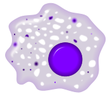 | 20〜50µm[6] | 寿命を迎えた赤血球・白血球・血小板や裸核・各種細胞などの細胞残屑と病原体の食作用(抱き込みと消化)、およびリンパ球に対する抗原提示、リンパ球の刺激、骨髄において幼若な赤血球の育成など | 数日〜数箇月、ときには数年 | |||
| 樹状細胞 |  |  | Tリンパ球を動かす抗原提示 | マクロファージと同じ単球系細胞 |
注、画像の色および顆粒の色の説明はギムザ染色したものでの説明である、実際の血液中の白血球は無色半透明である。
好中球の異物貪食動画像
| |

[1] Neutrophils display highly directional amoeboid motility in infected footpad and phalanges. Intravital imaging was performed in the footpad path of LysM-eGFP mice 20 min after infection with LM. [12]
脚注
- ^ 例えば白血球増加症は実質的には好中球増加症である。
- ^ a b c 小川『内科学書』p.15
- ^ a b c d e f g h i j k l 日本検査血液学会編、スタンダード検査血液学第2版、医歯薬出版、2008、p.50
- ^ 高久史麿監修『臨床検査データブック 2003-2004』(医学書院、2003)、 p.307
- ^ 『臨床検査データブック 2003-2004』p.307
- ^ a b c d e 小川 哲平、大島 年照、浅野 茂隆編著、血液学、内外医学社、1991
- ^ Brinkmann V, et al. Neutrophil extracellular traps kill bacteria. Science 2004: 303; 1532-1535.
- ^ Zawrotniak M, et al. Neutrophil extracellular traps (NETs)-frmation and implications. Acta Biochim Pol 2013: 60; 277-284.
- ^ 小川『内科学書』p.18
- ^ a b 日本検査血液学会編、スタンダード検査血液学第2版、医歯薬出版、2008、p.48
- ^ a b 日本検査血液学会編、スタンダード検査血液学第2版、医歯薬出版、2008、p.47
- ^ Public Library of Science
参考文献
- 笹月 健彦 監訳『免疫生物学 原書第5版』南江堂 2003年 ISBN 9784524235223
- 浅野茂隆、池田康夫、内山卓 監修 『三輪血液病学』文光堂、2006年、ISBN 4-8306-1419-6
- 小川聡 総編集 『内科学書』Vol.6 改訂第7版、中山書店、2009年、ISBN 978-4-521-73173-5
- 小川 哲平、大島 年照、浅野 茂隆編著、『血液学』、中外医学社、1991年
- 日本検査血液学会 編集『スタンダード検査血液学』初版、医歯薬出版、2003年、ISBN 4-263-22271-7
関連項目
This article is taken from the Japanese Wikipedia White blood cell
This article is distributed by cc-by-sa or GFDL license in accordance with the provisions of Wikipedia.
In addition, Tranpedia is simply not responsible for any show is only by translating the writings of foreign licenses that are compatible with CC-BY-SA license information.
0 개의 댓글:
댓글 쓰기